Distraction Osteogenesis
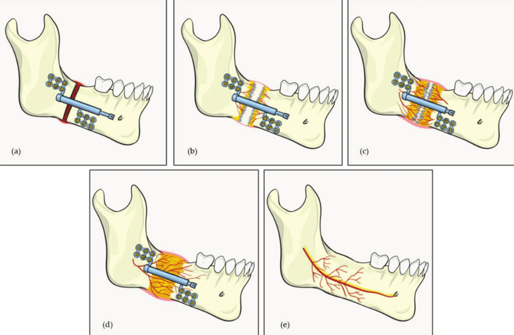
Distraction osteogenesis is a procedure utilized in orthopedic surgery to treat bone abnormalities. It is also known as callus distraction, allostasis, and osteodistraction. This surgical technique is commonly used to extend limbs. In other words, distraction osteogenesis is a technique to grow a long bone from a shorter one. Once a bone is cut during surgery, a device known as a distractor gradually pulls the two pieces of bone apart. The gradual separation of bones is not painful. Children claim that it hurts less than wearing braces to straighten their teeth. To bridge the gap, new bone emerges (osteogenesis). This occurs after your child has been discharged from the hospital and takes several months. Distraction osteogenesis allows for broader bone position improvements than are achievable with a single conventional surgery. This enhances the outcomes and could reduce the number of surgeries required by a child during their lifetime.
Since every distraction osteogenesis surgery is performed while the patient is under general anesthesia, there is no discomfort throughout the process. Following surgery, you will be provided analgesics (painkillers) to keep you comfortable and antibiotics to combat infection. Some patients may experience slight discomfort when the distraction device is applied to gradually separate the bones. Generally speaking, gradual movement of bone segments causes discomfort comparable to having braces tightened.
We have extensive expertise in treating distraction osteogenesis in children of various ages. Our patients include the following:
- Babies who have difficulty breathing
- Toddlers with a narrow jaw who require a breathing tube inside the windpipe
- Children whose cheekbones and eye sockets do not provide adequate protection for their eyes
- Teens with improperly aligned jaws
We customize treatment based on your child’s condition and their specific requirements. Our craniomaxillofacial surgeon uses 3-D imaging and state-of-the-art software to create detailed maps of your child’s face and skull during treatment planning. Our surgical expert can use 3-D imaging to estimate how movement in one area of the face would influence other parts. The software also takes into consideration the potential growth of the bone structure. This allows the surgeon to precisely determine how much and in which direction to move the various bones of the face.
